

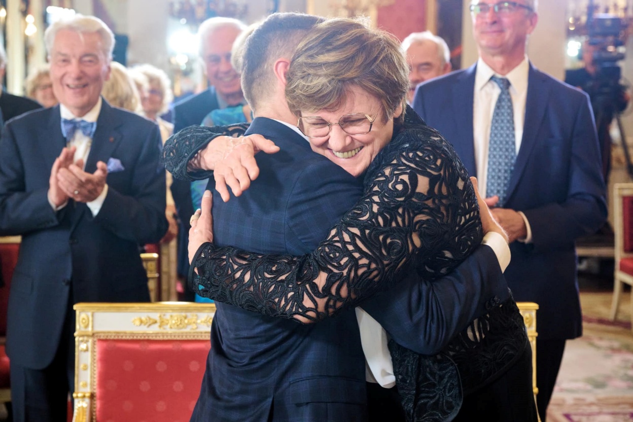
President Honors two of the great Hungarian scientists of our time.Continue reading

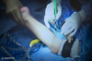
For the first time, plastic surgeons and ophthalmologists from Semmelweis University performed a pioneering surgery together. During this procedure, a 15-centimeter nerve was removed from the patient’s leg and implanted between a sensory nerve and the cornea of the eye, enabling the patient to regain sensory function in the cornea.
Conditions such as diabetes, tumors, nerve issues, facial nerve palsies, viral infections, sclerosis, and congenital brain diseases can cause corneal nerve damage, leading to loss of sensation on the eye surface. This results in impaired protective mechanisms, such as the inability to automatically tear or close the eye when exposed to foreign objects.
Dr. Zoltán Klárik, a plastic surgeon at the Department of Surgery, Transplantation, and Gastroenterology, developed this specialized procedure at Semmelweis University.
The technique was presented at the annual conference of the American Society for Reconstructive Microsurgery.
Photo: Semmelweis Egyetem / Zellei Boglárka
According to Dr. Klárik, “This procedure is used for conditions where lack of sensitivity on the eye’s surface prevents the patient from sensing damage to the cornea. This inadequate defense mechanism can lead to loss of the blink reflex, insufficient tear distribution, constant eye irritation, and reduced production of nerve-derived proteins crucial for maintaining and repairing the corneal epithelium. Ultimately, this can result in corneal ulcers and potential blindness.”
The inaugural surgery was performed on a middle-aged woman by a team of surgeons and ophthalmologists. During the procedure, a 15-centimeter nerve fiber was extracted from her leg without compromising leg function. “My role involved dividing the thick leg nerve into multiple fibers, suturing them under a microscope, and then passing the nerve through a subcutaneous tunnel system on the forehead from the intact side,” explained Dr. Klárik.
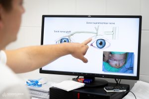
Photo: Semmelweis Egyetem / Zellei Boglárka
Dr. Ágnes Füst, leading the ophthalmology team, subsequently guided the nerve through the eyelid to the ocular surface, threading it under the conjunctiva around the cornea and securing it at five points. This meticulous ophthalmic and plastic microsurgery involved suturing millimeter-thick structures under an operating microscope. Dr. Füst recalled,
the most challenging aspect of the operation was coordinating the sequence of steps between the plastic surgeon and the ophthalmology team.”
The patient had been suffering from recurrent, non-healing corneal ulcers due to nerve damage caused by a central nervous system tumor. Prior to this surgery, she had undergone three eye surgeries in attempts to heal the ulcers and preserve vision. Since the specialized procedure, however, the ulcer has not recurred, and the patient’s quality of life has significantly improved, averting the risk of complete vision loss.
Via semmelweis.hu; Featured Image: Pixabay